Magnesium ferrite nanoparticles as cancer treatment adjuvants: synthesis, characterization, and heating capacity
Keywords:
Ferrites, Hyperthermia, Microemulsion, Nanoparticles, Superparamagnetic.Abstract
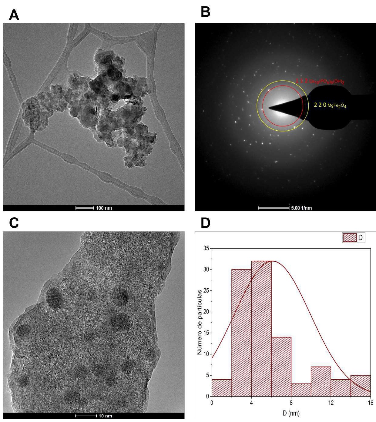
A microemulsion wet synthesis method was employed to obtain magnetic nanoparticles with the general formula MgxFe3-xO4, where x = 0.2, 0.4, 0.6, 0.8, and 1. After evaluating the magnetic properties and heating capacity of the synthesized powders, it was found that the most suitable results correspond to the values of x = 0.8 and 1. XRD analysis revealed a spinel structure corresponding to the pure phase of MgFe2O4, formed due to magnesium metal cations (Mg2+) replacing iron metal ions (Fe3+ - Fe2+). Using the SEM-EDX technique, aggregates of nanoparticles ranging from 5 to 20 µm were found (heat treatment at 900 °C). The results of the TGA-DSC analysis indicated a 50% loss of the precursor material and showed the onset of crystalline phase formation at 500 °C. TEM analysis confirms the presence of the MgFe2O4 phase, with a spherical morphology and an average particle size of 31 nm. VSM analysis demonstrated superparamagnetic behavior, where the maximum saturation magnetization value found in the nanoparticles (MgFe2O4) was 18.52 emu/g. The solid-state magnetic induction test revealed that using 6.0 mg of nanoparticles per ml of solution allows the temperature of the medium (H2O) to rise to 44.32 °C in less than 10 minutes. Cytotoxicity levels for erythrocyte cells in the presence of magnesium ferrites were up to 60 hours higher than 50% and very close to the control used. Based on the results, these nanoparticles could be effective in cancer treatment through magnetic hyperthermia therapy, as they can reach temperatures that induce thermal instability in proteins and nutrients in the affected area, leading to the death of neoplastic cells. Unlike normal cells, cancer cells exhibit greater sensitivity to temperature variations.
References
[1] A. R. K. Santhiya R, “Role of metal oxide ferrites in the process of magnetic hyperthermia”, J. Therm. Biol., 2024.
[2] P. S. Mkwae et al., “Insightful acetone gas sensing behaviour of Ce substituted MgFe2O4 spinel nano-ferrites”, J. Mater. Res. Technol., vol. 9, no. 6, pp. 16252–16269, 2020, doi: 10.1016/j.jmrt.2020.11.079.
[3] A. M. Jumaa, M.B., Mubarak, T.H. & Mohammad, “Exploring Cu-substituted Zn nanoferrites: synthesis, structural, magnetic, morphological, and antibacterial properties”, J Sol-Gel Sci Technol, 2022, [Online]. Available: https://doi.org/10.1007/s10971-025-06742-4.
[4] G. M. M. Frausto Gascón, Juan Salvador, Alexa Yael García-Fimbres, Celia Paola Gutierrez Aranda, Ximena Dannae Hernández Muñoz, Paola OrozcoF ernández, Teresa Alejandra Razo Lazcano, “Síntesis De nanopartículas De Oro Utilizando Nano-Emulsiones Y Extractos Naturales Como Agentes Reductores”, Jovenes en la Cienc. 21, 2023, [Online]. Available: https://www.jovenesenlaciencia.ugto.mx/index.php/jovenesenlaciencia/article/view/3943.
[5] D. A. B. Chetan Pandit, Arpita Roy, Suresh Ghotekar, Ameer Khusro, Mohammad Nazmul Islam, Talha Bin Emran, Siok Ee Lam, Mayeen Uddin Khandaker, “Biological agents for synthesis of nanoparticles and their applications”, J. King Saud Univ. - Sci., vol. 101869, no. ISSN 1018-3647, 2022, doi: https://doi.org/10.1016/j.jksus.2022.101869.
[6] D. et al. Malik, A.Q., Mir, T.u.G., Kumar, “A review on the green synthesis of nanoparticles, their biological applications, and photocatalytic efficiency against environmental toxins”, Env. Sci Pollut Res 30, vol. 69796–6982, 2023, doi: https://doi.org/10.1007/s11356-023-27437-9.
[7] H. H. and Z. G. Zhang D, Ma X-l, Gu Y, “Green Synthesis of Metallic Nanoparticles and Their Potential Applications to Treat Cancer”, Front. Chem, vol. 8, 2020, doi: https://doi.org/10.3389/fchem.2020.00799.
[8] C. Guyton and J. Hall, Guyton Y Hall Tratado De Fisiología Médica John E. Hall, vol. 14a. 2021.
[9] T. Pulingam, P. Foroozandeh, J. Chuah, and K. Sudesh, “Exploring Various Techniques for the Chemical and Biological Synthesis of Polymeric Nanoparticles”, 2022, doi: DOI:10.3390/nano12030576.
[10] Ana Ketzaly Calvillo Anguiano, “Evaluación de la viabilidad celular y la actividad antimicrobiana de nanopartículas de cobre Tesis”. 2019, [Online]. Available: http://www.fc.uaslp.mx/pca/tesis/2019Maestria/CalvilloAnguianoAnaKetzaly-Maestria201989.pdf.
[11] Z. V. Cancino J, Marangoni V.S, “Nanomedicine, Nanoscience and its applications”, Elsevier, pp. 71–84, 2017, doi: https://doi.org/10.1016/B978-0-323-49780-0.00003-X.
[12] F. A. S. da Silva and M. F. de Campos, “Study of heating curves generated by magnetite nanoparticles aiming application in magnetic hyperthermia”, Brazilian J. Chem. Eng., vol. 37, no. 3, pp. 543–553, 2020, doi: 10.1007/s43153-020-00063-5.
[13] J. K. Man Shu, Jingguang Wang, Ziyang Xu, Teliang Lu, Yue He, Renshan Li, Guoqing Zhong, Yunbo Yan, Yu Zhang, Xiao Chu, “Targeting nanoplatform synergistic glutathione depletion-enhanced chemodynamic, microwave dynamic, and selective-microwave thermal to treat lung cancer bone metastasis”, ioactive Mater., vol. 39, pp. 544–561, 2024, doi: https://doi.org/10.1016/j.bioactmat.2024.04.016.
[14] Y. Lu, W. Dong, J. Ding, W. Wang, and A. Wang, “Hydroxyapatite Nanomaterials: Synthesis, Properties, and Functional Applications”, Nanomater. from Clay Miner., pp. 485–536, 2019, doi: 10.1016/B978-0-12-814533-3.00010-7.
[15] A. Ravi, Rangnath, Mishra and S. Anamika, Ahmad, “Fabrication of Superparamagnetic Bimetallic Magnesium Nanoferrite Using Green Polyol: Characterization and Anticancer Analysis in Vitro on Lung Cancer Cell Line A549”, Am. Chem. Soc., vol. 5, 2022, doi: doi: 10.1021/acsabm.2c0072910.1021/acsabm.2c00729.
[16] M. G. & J. J. Souhir Bouzidi, Najah Rhimi, Jamila Dhahri, Zouhaier Aloui, Abdelaziz Bouazizi, Kamel Khirouni, “Eu-doped Mg–Ni ferrites: a reliable strategy to enhance dielectric properties for high-frequency applications”, J Mater Sci Mater Electron, vol. 36, p. 32, 2025, doi: https://doi.org/10.1007/s10854-024-14058-2.
[17] G. Orhan, Z., Daş, E. & Bozkurt, “Microemulsion synthesis of SnO2 nanoparticles and their integration in Au/n-Si/Al device structure”, J Mater Sci Mater Electron, vol. 36, p. 158, 2025, doi: https://doi.org/10.1007/s10854-025-14242-y.
[18] M. Tadic, J. Lazovic, M. Panjan, and S. Kralj, “Hierarchical iron oxide nanocomposite: Bundle-like morphology, magnetic properties and potential biomedical application”, Ceram. Int., vol. 48, no. 11, pp. 16015–16022, 2022, doi: 10.1016/j.ceramint.2022.02.145.
[19] M. Junaid et al., “Structural , spectral , dielectric and magnetic properties of indium substituted copper spinel ferrites synthesized via sol gel technique”, Ceram. Int., no. June, pp. 1–9, 2020, doi: 10.1016/j.ceramint.2020.07.227.
[20] A. B. K. Arun V. Bagade, Sangita N. Pund, Pratik A. Nagwade, Brajesh Kumar, Satish U. Deshmukh, “Ni-doped Mg-Zn nano-ferrites: Fabrication, characterization, and visible-light-driven photocatalytic degradation of model textile dyes”, Catal. Commun., vol. 181, 2023, doi: https://doi.org/10.1016/j.catcom.2023.106719.
[21] M. D. S. Moya Carlos Ferrer Giménez, Vicente Amigó Borrás, “Fundamentos de ciencia de los materiales”, Universidad politecnica de Valencia. 2023, [Online]. Available: https://www.upv.es/materiales/Fcm/Indice_FCM.html.
[22] S. Tong, H. Zhu, and G. Bao, “Magnetic iron oxide nanoparticles for disease detection and therapy”, Mater. Today, vol. 31, no. xx, pp. 86–99, 2019, doi: 10.1016/j.mattod.2019.06.003.
[23] M. Tadic, S. Kralj, and L. Kopanja, “Synthesis, particle shape characterization, magnetic properties and surface modification of superparamagnetic iron oxide nanochains”, Mater. Charact., vol. 148, no. December 2018, pp. 123–133, 2019, doi: 10.1016/j.matchar.2018.12.014.
[24] L. Kopanja, M. Tadić, S. Kralj, and J. Žunić, Shape and aspect ratio analysis of anisotropic magnetic nanochains based on TEM micrographs, vol. 44, no. 11. Elsevier Ltd and Techna Group S.r.l., 2018.
[25] T. Tatarchuk et al., “Magnesium-zinc ferrites as magnetic adsorbents for Cr(VI) and Ni(II) ions removal: Cation distribution and antistructure modeling”, Chemosphere, vol. 270, 2021, doi: 10.1016/j.chemosphere.2020.129414.
[26] J. Uddin and Y. Jeong, “Chemosphere Application of magnesium ferrite nanomaterials for adsorptive removal of arsenic from water : Effects of Mg and Fe ratio”, Chemosphere, vol. 307, no. P3, p. 135817, 2022, doi: 10.1016/j.chemosphere.2022.135817.
[27] H. Z. Feng Ding, Guobin Qiu, Fengliang Tan, Tengyan Wu, Longzhen Ding, Xin Liu, “Preparation and characterization of PEGylated magnetic ferrite with a biocompatible delivery for DOX”, Appl. Phys. A, vol. 12, p. 129, 2023, doi: https://doi.org/10.1007/s00339-023-07160-5.
[28] V. Vilas-Boas, F. Carvalho, and B. Espiña, “Magnetic hyperthermia for cancer treatment: Main parameters affecting the outcome of in vitro and in vivo studies”, Molecules, vol. 25, no. 12, pp. 1–30, 2020, doi: 10.3390/molecules25122874.
[29] B. D. Cardoso et al., “Magnetoliposomes based on shape anisotropic calcium/magnesium ferrite nanoparticles as nanocarriers for doxorubicin”, Pharmaceutics, vol. 13, no. 8, 2021, doi: 10.3390/pharmaceutics13081248.
[30] G. Nandhini and M. K. Shobana, “Efecto de la moringa oleifera con nanoferritas de Mn y Ni puras y dopadas con Zn para aplicaciones de hipertermia,” Mater. Today Commun., vol. 39, 2024, doi: https://doi.org/10.1016/j.mtcomm.2024.109046.
[31] Marta Vassallo, Daniele Martella, Gabriele Barrera, Federica Celegato, Marco Coïsson, Ricardo Ferrero, Elena S. Olivetti, Adriano Tróia, Hüseyin Sözeri, Camilla Parmeggiani, Diederik S. Wiersma, Paola Tiberto and A. Manzin, “Mejora de las propiedades de hipertermia de las nanopartículas de óxido de hierro mediante recubrimiento superficial”, ACS Omega, vol. 8, 2023, [Online]. Available: https://pubs-acs-org.translate.goog/doi/10.1021/acsomega.2c06244?_x_tr_sl=en&_x_tr_tl=es&_x_tr_hl=es&_x_tr_pto=tc.
[32] S. Kanagesan et al., “Evaluation of antioxidant and cytotoxicity activities of copper ferrite (CuFe2O4) and zinc ferrite (ZnFe2O4) nanoparticles synthesized by sol-gel self-combustion method”, Appl. Sci., vol. 6, no. 9, pp. 1–13, 2016, doi: 10.3390/app6090184.
[33] A. Rafieepour et al., “Investigation of the effect of magnetite iron oxide particles size on cytotoxicity in A549 cell line”, Toxicol. Ind. Health, vol. 35, no. 11–12, pp. 703–713, 2019, doi: 10.1177/0748233719888077.
[34] K. Shahzad et al., “Effect of Magnesium Substitution on Structural, Magnetic and Biological Activity of Co(1-x)Mg(x)Fe2O4 Nano-colloids”, J. Clust. Sci., vol. 32, no. 4, pp. 1003–1014, 2021, doi: 10.1007/s10876-020-01862-z.