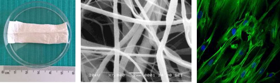
Assessment of the cytotoxicity of PCL and PLGA scaffolds with dental pulp stem cells for tissue engineering
Keywords:
Dental pulp stem cells, lactic-co-glycolic acid, poly - ε caprolactone, scaffolds, tissue engineering.Abstract
Materials of different nature such as poly - ε caprolactone and glycolic poly-lactic acid, have been used in regenerative medicine with human applications and dental pulp stem cells represent an attractive source of cells to be used in in vitro assays due to their easy accessibility, good cell viability and the fact that they are obtained from human third molars in a routine extraction.
Objective: To evaluate the in vitro cytotoxicity of PCL/PLGA scaffolds cultured with dental pulp stem cells.
Materials and methods: An experimental in vitro study was carried out evaluating PCL/ PLGA scaffolds in two portions (80:20 and 90:10, respectively) on which DPSC obtained from third molars were planted. The cell viability was evaluated at three times 3, 7 and 10 days by the MTT assay. Data analysis was performed with the Stata software version 14 and the Student's "t" test.
Results: Culture and characterization of dental pulp stem cells were positive for CD44 and Stro-1, the results show an increase in cell viability from day 3 in the control groups and both polymer mixtures. But there was a lower metabolic activity in the PLGA (90:10) compared to the PLC/PLGA (80:20).
Conclusions: PCL/PLGA 80:20 scaffolds showed greater cell viability at days 7 and 10 compared to the PCL/PLGA 90:10. It was also shown that neither mixture has cytotoxic effect on dental pulp stem cells.
References
1. Berthiaume, F., Maguire, T. J., & Yarmush, M. L. (2011). Tissue engineering and regenerative medicine: history, progress, and challenges. Annual review of chemical and biomolecular engineering, 2, 403–430. https://doi.org/10.1146/annurev-chembioeng-061010-114257.
2. Pittenger, M. F., Mackay, A. M., Beck, S. C., Jaiswal, R. K., Douglas, R., Mosca, J. D., Moorman, M. A., Simonetti, D. W., Craig, S., & Marshak, D. R. (1999). Multilineage potential of adult human mesenchymal stem cells. Science (New York, N.Y.), 284(5411), 143–147. https://doi.org/10.1126/science.284.5411.143.
3. Caplan, A. I. (1990). Cell delivery and tissue regeneration. Journal of Controlled Release, 11(1-3), 157–165. https://doi.org/10.1016/0168-3659(90)90129-H
4. Ullah, I., Subbarao, R. B., & Rho, G. J. (2015). Human mesenchymal stem cells - current trends and future prospective. Bioscience reports, 35(2), e00191. https://doi.org/10.1042/BSR20150025.
5. Friedenstein, A. J., Deriglasova, U. F., Kulagina, N. N., Panasuk, A. F., Rudakowa, S. F., Luriá, E. A., & Ruadkow, I. A. (1974). Precursors for fibroblasts in different populations of hematopoietic cells as detected by the in vitro colony assay method. Experimental hematology, 2(2), 83–92.
6. Erices, A., Conget, P., & Minguell, J. J. (2000). Mesenchymal progenitor cells in human umbilical cord blood. British journal of haematology, 109(1), 235–242. https://doi.org/10.1046/j.1365-2141.2000.01986.x.
7. Gronthos, S., Mankani, M., Brahim, J., Robey, P. G., & Shi, S. (2000). Postnatal human dental pulp stem cells (DPSCs) in vitro and in vivo. Proceedings of the National Academy of Sciences of the United States of America, 97(25), 13625–13630. https://doi.org/10.1073/pnas.240309797.
8. Sonoyama, W., Liu, Y., Fang, D., Yamaza, T., Seo, B. M., Zhang, C., Liu, H., Gronthos, S., Wang, C. Y., Wang, S., & Shi, S. (2006). Mesenchymal stem cell-mediated functional tooth regeneration in swine. PloS one, 1(1), e79. https://doi.org/10.1371/journal.pone.0000079.
9. Sasaki, R., Aoki, S., Yamato, M., Uchiyama, H., Wada, K., Ogiuchi, H., Okano, T., & Ando, T. (2011). PLGA artificial nerve conduits with dental pulp cells promote facial nerve regeneration. Journal of tissue engineering and regenerative medicine, 5(10), 823–830. https://doi.org/10.1002/term.387.
10. Tamaki, Y., Nakahara, T., Ishikawa, H., & Sato, S. (2013). In vitro analysis of mesenchymal stem cells derived from human teeth and bone marrow. Odontology, 101(2), 121–132. https://doi.org/10.1007/s10266-012-0075-0.
11. Bártolo, P.J., Chua, C.K., Almeida, H.A., Chou S.M. y Lim S.C. (2009). Biomanufacturing for tissue engineering: Present and future trends, Virtual and Physical Prototyping, 4:4, 203-216. https://doi.org/10.1080/17452750903476288.
12. Bartolo, P., Kruth, J.P., Silva, J., Levy G., Malshe, A. Rajurkar, K.P., Mitsuishi, M., Ciurana, J. y Leu M. (2012). Biomedical production of implants by additive electro-chemical and physical processes. CIRP annals 2012; 61(2): 635-655. https://doi.org/10.1016/j.cirp.2012.05.005.
13. Domingos, M., Chiellini, F., Gloria, A., Ambrosio, L., Bartolo, P. and Chiellini, E. (2012), "Effect of process parameters on the morphological and mechanical properties of 3D Bioextruded poly(ε‐caprolactone) scaffolds", Rapid Prototyping Journal, Vol. 18 No. 1, pp. 56-67. https://doi.org/10.1108/13552541211193502.
14. De Santis, R., Gloria, A., Russo, T., D’Amora, U., Zeppetelli, S., Tampieri, A., Herrmannsdörfer T. y Ambrosio, L. A route toward the development of 3D magnetic scaffolds with tailored mechanical and morphological properties for hard tissue regeneration: Preliminary study: A basic approach toward the design of 3D rapid prototyped magnetic scaffolds for hard-tissue regeneration is presented and validated in this paper. Virtual and Physical Prototyping 2011; 6(4): 189-195. https://doi.org/10.1080/17452759.2011.631324.
15. Collins, M. N., & Birkinshaw, C. (2013). Hyaluronic acid based scaffolds for tissue engineering--a review. Carbohydrate polymers, 92(2), 1262–1279. https://doi.org/10.1016/j.carbpol.2012.10.028
16. Afewerki, S., Sheikhi, A., Kannan, S., Ahadian, S., & Khademhosseini, A. (2018). Gelatin-polysaccharide composite scaffolds for 3D cell culture and tissue engineering: Towards natural therapeutics. Bioengineering & translational medicine, 4(1), 96–115. https://doi.org/10.1002/btm2.10124
17. Badylak, S. F., Freytes, D. O., & Gilbert, T. W. (2009). Extracellular matrix as a biological scaffold material: Structure and function. Acta biomaterialia, 5(1), 1–13. https://doi.org/10.1016/j.actbio.2008.09.013.
18. Patrício, T., Domingos, M., Gloria, A., & Bártolo, P. (2013). Characterisation of PCL and PCL/PLA Scaffolds for Tissue Engineering. Procedia CIRP, 5, 110–114. https://doi.org/10.1016/j.procir.2013.01.022.
19. Nitya, G., Nair, G. T., Mony, U., Chennazhi, K. P., & Nair, S. V. (2012). In vitro evaluation of electrospun PCL/nanoclay composite scaffold for bone tissue engineering. Journal of materials science. Materials in medicine, 23(7), 1749–1761. https://doi.org/10.1007/s10856-012-4647-x.
20. Fu, W., Liu, Z., Feng, B., Hu, R., He, X., Wang, H., Yin, M., Huang, H., Zhang, H., & Wang, W. (2014). Electrospun gelatin/PCL and collagen/PLCL scaffolds for vascular tissue engineering. International journal of nanomedicine, 9, 2335–2344. https://doi.org/10.2147/IJN.S61375.
21. Zamani, F., Amani-Tehran, M., Latifi, M., & Shokrgozar, M. A. (2013). The influence of surface nanoroughness of electrospun PLGA nanofibrous scaffold on nerve cell adhesion and proliferation. Journal of materials science. Materials in medicine, 24(6), 1551–1560. https://doi.org/10.1007/s10856-013-4905-6.
22. García, N.G. (2013). Electrospinning: una técnica fascinante para la obtención de nanofibras poliméricas. Revista de plásticos modernos: Ciencia y tecnología de polímeros; 677: 166-173.
23. O'Driscoll S. W. (1999). Articular cartilage regeneration using periosteum. Clinical orthopaedics and related research, (367 Suppl), S186–S203. https://doi.org/10.1097/00003086-199910001-00020.
24. Majumdar, M. K., Banks, V., Peluso, D. P., & Morris, E. A. (2000). Isolation, characterization, and chondrogenic potential of human bone marrow-derived multipotential stromal cells. Journal of cellular physiology, 185(1), 98–106. https://doi.org/10.1002/1097-4652(200010)185:1<98::AID-JCP9>3.0.CO;2-1.
25. Hiep, N. T., & Lee, B. T. (2010). Electro-spinning of PLGA/PCL blends for tissue engineering and their biocompatibility. Journal of materials science. Materials in medicine, 21(6), 1969–1978. https://doi.org/10.1007/s10856-010-4048-y
26. Milosevic, M., Stojanovic, D. B., Simic, V., Grkovic, M., Bjelovic, M., Uskokovic, P. S., & Kojic, M. (2020). Preparation and modeling of three-layered PCL/PLGA/PCL fibrous scaffolds for prolonged drug release. Scientific reports, 10(1), 11126. https://doi.org/10.1038/s41598-020-68117-9
27. Maurmann N., D. P Pereira, D. Burguez, F.D.A. de S Pereira, P. Inforçatti Neto, R. A. Rezende, D. Gamba, JVL da Silva y P. Pranke. (2017). Mesenchymal stem cells cultivated on scaffolds formed by 3D printed PCL matrices, coated with PLGA electrospun nanofibers for use in tissue engineering. Biomed. Phys. Eng. Express 3,045005. https://doi.org/10.1088/2057-1976/aa6308.
28. Sánchez-Pech, J. C., Rosales-Ibáñes, R., Cauich-Rodriguez, J. V., Carrillo-Escalante, H. J., Rodríguez-Navarrete, A., Avila-Ortega, A., & Hernández-Sánchez, F. (2020). Design, synthesis, characterization, and cytotoxicity of PCL/PLGA scaffolds through plasma treatment in the presence of pyrrole for possible use in urethral tissue engineering. Journal of biomaterials applications, 34(6), 840–850. https://doi.org/10.1177/0885328219882638.
29. Guzmán-Uribe, D., Estrada, K. N., Guillén, A., Pérez, S. M., & Ibáñez, R. R. (2012). Development of a three-dimensional tissue construct from dental human ectomesenchymal stem cells: in vitro and in vivo study. The open dentistry journal, 6, 226–234. https://doi.org/10.2174/1874210601206010226.
30. Flores-Cedillo, M. L., Alvarado-Estrada, K. N., Pozos-Guillén, A. J., Murguía-Ibarra, J. S., Vidal, M. A., Cervantes-Uc, J. M., Rosales-Ibáñez, R., & Cauich-Rodríguez, J. V. (2016). Multiwall carbon nanotubes/polycaprolactone scaffolds seeded with human dental pulp stem cells for bone tissue regeneration. Journal of materials science. Materials in medicine, 27(2), 35. https://doi.org/10.1007/s10856-015-5640-y.
31. Martin-Del-Campo, M., Rosales-Ibañez, R., Alvarado, K., Sampedro, J. G., Garcia-Sepulveda, C. A., Deb, S., San Román, J., & Rojo, L. (2016). Strontium folate loaded biohybrid scaffolds seeded with dental pulp stem cells induce in vivo bone regeneration in critical sized defects. Biomaterials science, 4(11), 1596–1604. https://doi.org/10.1039/c6bm00459h.3.
32. Dominici, M., Le Blanc, K., Mueller, I., Slaper-Cortenbach, I., Marini, F., Krause, D., Deans, R., Keating, A., Prockop, D.j, & Horwitz, E. (2006). Minimal criteria for defining multipotent mesenchymal stromal cells. The International Society for Cellular Therapy position statement. Cytotherapy, 8(4), 315–317. https://doi.org/10.1080/14653240600855905
33. Sivasankar, V., & Ranganathan, K. (2016). Growth characteristics and expression of CD73 and CD146 in cells cultured from dental pulp. Journal of investigative and clinical dentistry, 7(3), 278–285. https://doi.org/10.1111/jicd.12155.
34. Tang, Z. G., Callaghan, J. T., & Hunt, J. A. (2005). The physical properties and response of osteoblasts to solution cast films of PLGA doped polycaprolactone. Biomaterials, 26(33), 6618–6624. https://doi.org/10.1016/j.biomaterials.2005.04.013
35. Kim, J. Y., & Cho, D.-W. (2009). Blended PCL/PLGA scaffold fabrication using multi-head deposition system. Microelectronic Engineering, 86(4-6), 1447–1450.https://doi.org/10.1016/j.mee.2008.11.026
36. Calvert, J. W., Marra, K. G., Cook, L., Kumta, P. N., DiMilla, P. A., & Weiss, L. E. (2000). Characterization of osteoblast-like behavior of cultured bone marrow stromal cells on various polymer surfaces. Journal of biomedical materials research, 52(2), 279–284. https://doi.org/10.1002/1097-4636(200011)52:2<279::aid-jbm6>3.0.co;2-8
37. Elder, S., Roberson, J. G., Warren, J., Lawson, R., Young, D., Stokes, S., & Ross, M. K. (2022). Evaluation of Electrospun PCL-PLGA for Sustained Delivery of Kartogenin. Molecules (Basel, Switzerland), 27(12), 3739. https://doi.org/10.3390/molecules27123739
38.Fioretta, E. S., Simonet, M., Smits, A. I., Baaijens, F. P., & Bouten, C. V. (2014). Differential response of endothelial and endothelial colony forming cells on electrospun scaffolds with distinct microfiber diameters. Biomacromolecules, 15(3), 821–829. https://doi.org/10.1021/bm4016418.
39. Nezarati, R. M., Eifert, M. B., & Cosgriff-Hernandez, E. (2013). Effects of humidity and solution viscosity on electrospun fiber morphology. Tissue engineering. Part C, Methods, 19(10), 810–819. https://doi.org/10.1089/ten.TEC.2012.0671.
40. Rosales-Ibáñez, R., Cubo-Mateo, N., Rodríguez-Navarrete, A., González-González, A. M., Villamar-Duque, T. E., Flores-Sánchez, L. O., & Rodríguez-Lorenzo, L. M. (2021). Assessment of a PCL-3D Printing-Dental Pulp Stem Cells Triplet for Bone Engineering: An In Vitro Study. Polymers, 13(7), 1154. https://doi.org/10.3390/polym13071154
41.Alraies, A., Alaidaroos, N. Y., Waddington, R. J., Moseley, R., & Sloan, A. J. (2017). Variation in human dental pulp stem cell ageing profiles reflect contrasting proliferative and regenerative capabilities. BMC cell biology, 18(1), 12. https://doi.org/10.1186/s12860-017-0128-x
42. Amid, R., Kadkhodazadeh, M., Enssi, M., & Dehanvi, F. (2021). In Vitro Activity of Dental Pulp Stem Cells versus the Bone Marrow Stem Cells Cultured in Presence of a Bone Allograft. Journal of long-term effects of medical implants, 31(1), 7–14. https://doi.org/10.1615/JLongTermEffMedImplants.2020036956
43. Labedz-Maslowska, A., Bryniarska, N., Kubiak, A., Kaczmarzyk, T., Sekula-Stryjewska, M., Noga, S., Boruczkowski, D., Madeja, Z., & Zuba-Surma, E. (2020). Multilineage Differentiation Potential of Human Dental Pulp Stem Cells-Impact of 3D and Hypoxic Environment on Osteogenesis In Vitro. International journal of molecular sciences, 21(17), 6172. https://doi.org/10.3390/ijms21176172
44. Hynes R. O. (2009). The extracellular matrix: not just pretty fibrils. Science (New York, N.Y.), 326(5957), 1216–1219. https://doi.org/10.1126/science.1176009.
45. Ling, L. E., Feng, L., Liu, H. C., Wang, D. S., Shi, Z. P., Wang, J. C., Luo, W., & Lv, Y. (2015). The effect of calcium phosphate composite scaffolds on the osteogenic differentiation of rabbit dental pulp stem cells. Journal of biomedical materials research. Part A, 103(5), 1732–1745. https://doi.org/10.1002/jbm.a.35303.3.
46. Jiménez, N. T., Carlos Munévar, J., González, J. M., Infante, C., & Lara, S. (2018). In vitro response of dental pulp stem cells in 3D scaffolds: A regenerative bone material. Heliyon, 4(9), e00775. https://doi.org/10.1016/j.heliyon.2018.e00775.
47. Markovic, D., Karadzic, I., Jokanovic, V., Vukovic, A., Vucic, V. (2016). Biological aspects of application of nanomaterials in tissue engineering. Chem. Ind. Chem. Eng. Q. 22 (2) 145−153 https://doi.org/10.2298/CICEQ141231028M.
48. Chen, G., Chen, J., Yang, B., Li, L., Luo, X., Zhang, X., Feng, L., Jiang, Z., Yu, M., Guo, W., & Tian, W. (2015). Combination of aligned PLGA/Gelatin electrospun sheets, native dental pulp extracellular matrix and treated dentin matrix as substrates for tooth root regeneration. Biomaterials, 52, 56–70. https://doi.org/10.1016/j.biomaterials.2015.02.011.
49. Rickert, D., Lendlein, A., Peters, I., Moses, M. A., & Franke, R. P. (2006). Biocompatibility testing of novel multifunctional polymeric biomaterials for tissue engineering applications in head and neck surgery: an overview. European archives of oto-rhino-laryngology : official journal of the European Federation of Oto-Rhino-Laryngological Societies (EUFOS) : affiliated with the German Society for Oto-Rhino-Laryngology - Head and Neck Surgery, 263(3), 215–222. https://doi.org/10.1007/s00405-005-0950-1
50. Zou, H., Wang, G., Song, F. y Shi, X. (2017). Investigation of Human Dental Pulp Cells on a Potential Injectable Poly(lactic-co-glycolic acid) Microsphere Scaffold. J Endod; 43(5): 745-750. https://doi.org/10.1016/j.joen.2016.12.019.
51. Khojasteh, A., Motamedian, S. R., Rad, M. R., Shahriari, M. H., & Nadjmi, N. (2015). Polymeric vs hydroxyapatite-based scaffolds on dental pulp stem cell proliferation and differentiation. World journal of stem cells, 7(10), 1215–1221. https://doi.org/10.4252/wjsc.v7.i10.1215.
52. D'Souza, S., Faraj, J. A., Dorati, R., & DeLuca, P. P. (2014). A short term quality control tool for biodegradable microspheres. AAPS PharmSciTech, 15(3), 530–541. https://doi.org/10.1208/s12249-013-0052-0.
53. Gangolli, R. A., Devlin, S. M., Gerstenhaber, J. A., Lelkes, P. I., & Yang, M. (2019). A Bilayered Poly (Lactic-Co-Glycolic Acid) Scaffold Provides Differential Cues for the Differentiation of Dental Pulp Stem Cells. Tissue engineering. Part A, 25(3-4), 224–233. https://doi.org/10.1089/ten.TEA.2018.0041.
54. Atala, A., Kasper, F. K., & Mikos, A. G. (2012). Engineering complex tissues. Science translational medicine, 4(160), 160rv12. https://doi.org/10.1126/scitranslmed.3004890.