Synthesis of a 45S5 bioactive glass cementitious resin bonded composite material for orthodontic applications
Keywords:
Composite material, Bioactive glass, Cementitious resin, Orthodontics.Abstract
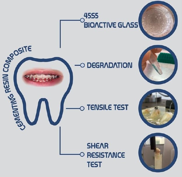
A composite material based on 45S5 bioactive glass and commercial cementitious resin for orthodontic applications, was obtained, taking two commercial resins as control. The bioactive glass was synthesized using sol-gel and stabilizing the dry gel using microwave assistance for 6 minutes, obtaining an amorphous material with good dimensional stability and transparency. A composite material was prepared using Transbond cementitious resin and 8% 45S5 bioactive glass. The resulting composite was subjected to degradation tests to determine the effect of artificial saliva on this material degradation over a period of one month. Sample composite degradation and the fluid pH variations were analyzed via SEM/EDS. Composite bioactivity was determined by exposing the material to simulated physiological fluid (SBF), showing the formation of biological apatite layers on the materials’ surface. The composite´s mechanical properties were analyzed by adhering the brackets with the composite material to previously preserved human dental pieces and the shear strength of the composite was determined along with the tension strength between the resin teeth interface. Analyzes were performed by triplicate. A Statistical ANOVA analysis was performed to determine significant mechanical test results. These data showed that the synthesized composite material exhibited better bioactivity, mechanical and degradational properties compared to the commercial resins.
References
Al, E., Adeyemi, A., Higham, S., Pender, Harrison, J. (2009). Factors affecting demineralization during orthodontic treatment: A post-hoc analysis of RCT recruits. Am. J. Orthod. Dentofac. Orthop., 139(2), 181–191. 10.1016/j.ajodo.2009.08.028
Seyhan-Cezairli, N., Serkan-Küçükekenci, A., & Başoğlu, H. (2019). Evaluation of shear bond strength between orthodontic brackets and three aged bulk fill composites. Odovtos International Journal of Dental Sciences, 21(3), 89-99. http://dx.doi.org/10.15517/ijds.v0i0.36005
Gao, Z., Chen, X., Wang, C., Song, J., Xu, J., Liu, X., ... & Suo, H. (2023). New strategies and mechanisms for targeting Streptococcus mutans biofilm formation to prevent dental caries: a review. Microbiological Research, 127526.
Zafar, M. S. (2020). Prosthodontic applications of polymethyl methacrylate (PMMA): An update. Polymers, 12(10), 2299. doi:10.3390/polym12102299
Vallittu, P., Närhi, P., Hupa L. (2015). Fiber glass-bioactive glass composite for bone replacing and bone anchoring implants. Dent. Mater., 31 (4), 371–381. 10.1016/j.dental.2015.01.003
DEGRAZIA, F.,LEITUNE,C., GARCIA, I., ARTHUR, R., SAMUEL, S., COLLARES, F. (2016) Effect of silver nanoparticles on the physicochemical and antimicrobial properties of an orthodontic adhesive. J. Appl. Oral Sci., 24(4), 404–410. 10.1590/1678-775720160154
Kim, H. J., Bae, H. E., Lee, J. E., Park, I. S., Kim, H. G., Kwon, J., & Kim, D. S. (2021). Effects of bioactive glass incorporation into glass ionomer cement on demineralized dentin. Scientific Reports, 11(1), 7016.
Manafi, S., Mirjalili, F., & Reshadi, R. (2019). Synthesis and evaluation of the bioactivity of fluorapatite–45S5 bioactive glass nanocomposite. Progress in biomaterials, 8(2), 77-89.
Pirayesh, H. (2010). Effects of manufacturing method on surface mineralization of bioactive glasses. University of Alberta, Edmonton, Alberta. https://doi.org/10.7939/R37G64
Kokubo T. and Takadama H. (2006). How useful is SBF in predicting in vivo bone bioactivity?. Biomaterials. 27(15), 2907–2915. https://doi.org/10.1016/j.biomaterials.2006.01.017
Vouzara, T., Roussou, K., Nikolaidis, A., Tolidis, K., Koulaouzidou, E. (2020). Organic eluates derived from intermediate restorative dental materials, Molecules, 25(7), 1–14. doi:10.3390/molecules25071593
Pulpdent Coporation. (2019). Safety data sheet: ACTIVA BioACTIVE-BASE/LINER.
Bauer J., Silva A., Moreno E., Campos P., Nunes C., Pigozzo A., Marins R. (2018). Dentin pretreatment with 45S5 and niobophosphate bioactive glass: Effects on pH, antibacterial, mechanical properties of the interface and microtensile bond strength, J. Mech. Behav. Biomed. Mater., 90, 374–380. https://doi.org/10.1016/j.jmbbm.2018.10.029
Barrios, C., Vila, V., Martinez, S., Encina A. (2017). Ph Salival como factor asociado a la caries dental, Rev. Fac. Odontol. Univ. Nac. (Cordoba)., 10(1), 13. https://core.ac.uk/reader/230848120
Silva, R. R. A., Marques, C. S., Arruda, T. R., Teixeira, S. C., & de Oliveira, T. V. (2023). Biodegradation of Polymers: Stages, Measurement, Standards and Prospects. Macromol, 3(2), 371-399. https://doi.org/ 10.3390/macromol3020023
Nawrocka, A., Nowak, J., Sauro, S., Hardan, L., Bourgi, R., & Lukomska-Szymanska, M. (2023). Shear Bond Strength of Metal and Ceramic Brackets Depending on Etching Protocol in Direct Bonding Technique. Materials, 16(20), 6697.
Skallevold, H., Rokaya, D., Khurshid, Z., Zafar, M. (2019). Bioactive glass applications in dentistry, Int. J. Mol. Sci., 20(23), 1–24. 10.3390/ijms20235960
Profeta A., Prucher, G. Bioactive-glass in periodontal surgery and implant dentistry, Dent. Mater. J., 34(5), 559–571. 10.4012/dmj.2014-233.
AlSamak, S., Alsaleem, N. R., & Ahmed, M. K. (2023). Evaluation of the shear bond strength and adhesive remnant index of color change, fluorescent, and conventional orthodontic adhesives: An in vitro study. International Orthodontics, 21(1), 100712. https://doi.org/10.1016/j.ortho.2022.100712
Álvarez-Monsiváis, M. E., Cruz-Ortiz, B. R., Olivas-Armendáriz, I., Sáenz-Galindo, A., Solís-Rosales, S. G., & Múzquiz-Ramos, E. M. (2022). Bioactividad y biocompatibilidad de compósitos de zirconia estabilizada con Y/hidroxiapatita-Cu, Fosfato Tricálcico-Ag o Fosfato Tricálcico-Ga. Afinidad, 79(596), 358-366. https://doi.org/10.55815/401133