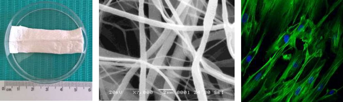
Evaluación de la citotoxicidad de andamios de PCL y PLGA con células troncales de pulpa dental para ingeniería tisular
Palabras clave:
Células troncales de pulpa dental, poli-ε-caprolactona. ácido poli-láctico-co-glicólico, andamios, ingeniería tisular.Resumen
Materiales de diferente naturaleza como poli- ε caprolactona y el ácido poli(láctico-co-glicólico), han sido empleados en medicina regenerativa con aplicaciones en humanos y, las células troncales de pulpa dental representan una fuente atractiva para emplearse en ensayos in vitro debido a su fácil acceso, buena viabilidad celular ya que se obtienen de terceros molares humanos en una extracción de rutina.
Objetivo: Evaluar la citotoxicidad in vitro de andamios de PCL/PLGA cultivados con células troncales de pulpa dental.
Materiales y métodos: Se realizó un estudio experimental in vitro evaluando los andamios de PCL/PLGA en dos proporciones (80:20 y 90:10, respectivamente) a los cuales se les sembraron células troncales de pulpa dental obtenidas de terceros molares. La viabilidad celular se evaluó a tres tiempos 3, 7 y 10 días mediante el ensayo MTT. El análisis de los datos se realizó con el software Stata versión 14 y la prueba "t" de Student.
Resultados: Se logró cultivar y caracterizar las células troncales de pulpa dental. Los resultados mostraron un aumento de la viabilidad celular a partir del día 3 en el grupo control y ambas mezclas de polímeros, pero existió una menor actividad metabólica en la mezcla PCL/PLGA (90:10) respecto a la mezcla PCL/PLGA (80:20).
Conclusiones: La mezcla PCL/PLGA 80:20 tuvo mayor viabilidad celular en los días 7 y 10 comparada con la PCL/PLGA de 90:10. También se demostró que ninguna de las dos mezclas tiene efectos citotóxicos en las células troncales de pulpa dental.
Referencias
1. Berthiaume, F., Maguire, T. J., & Yarmush, M. L. (2011). Tissue engineering and regenerative medicine: history, progress, and challenges. Annual review of chemical and biomolecular engineering, 2, 403–430. https://doi.org/10.1146/annurev-chembioeng-061010-114257.
2. Pittenger, M. F., Mackay, A. M., Beck, S. C., Jaiswal, R. K., Douglas, R., Mosca, J. D., Moorman, M. A., Simonetti, D. W., Craig, S., & Marshak, D. R. (1999). Multilineage potential of adult human mesenchymal stem cells. Science (New York, N.Y.), 284(5411), 143–147. https://doi.org/10.1126/science.284.5411.143.
3. Caplan, A. I. (1990). Cell delivery and tissue regeneration. Journal of Controlled Release, 11(1-3), 157–165. https://doi.org/10.1016/0168-3659(90)90129-H
4. Ullah, I., Subbarao, R. B., & Rho, G. J. (2015). Human mesenchymal stem cells - current trends and future prospective. Bioscience reports, 35(2), e00191. https://doi.org/10.1042/BSR20150025.
5. Friedenstein, A. J., Deriglasova, U. F., Kulagina, N. N., Panasuk, A. F., Rudakowa, S. F., Luriá, E. A., & Ruadkow, I. A. (1974). Precursors for fibroblasts in different populations of hematopoietic cells as detected by the in vitro colony assay method. Experimental hematology, 2(2), 83–92.
6. Erices, A., Conget, P., & Minguell, J. J. (2000). Mesenchymal progenitor cells in human umbilical cord blood. British journal of haematology, 109(1), 235–242. https://doi.org/10.1046/j.1365-2141.2000.01986.x.
7. Gronthos, S., Mankani, M., Brahim, J., Robey, P. G., & Shi, S. (2000). Postnatal human dental pulp stem cells (DPSCs) in vitro and in vivo. Proceedings of the National Academy of Sciences of the United States of America, 97(25), 13625–13630. https://doi.org/10.1073/pnas.240309797.
8. Sonoyama, W., Liu, Y., Fang, D., Yamaza, T., Seo, B. M., Zhang, C., Liu, H., Gronthos, S., Wang, C. Y., Wang, S., & Shi, S. (2006). Mesenchymal stem cell-mediated functional tooth regeneration in swine. PloS one, 1(1), e79. https://doi.org/10.1371/journal.pone.0000079.
9. Sasaki, R., Aoki, S., Yamato, M., Uchiyama, H., Wada, K., Ogiuchi, H., Okano, T., & Ando, T. (2011). PLGA artificial nerve conduits with dental pulp cells promote facial nerve regeneration. Journal of tissue engineering and regenerative medicine, 5(10), 823–830. https://doi.org/10.1002/term.387.
10. Tamaki, Y., Nakahara, T., Ishikawa, H., & Sato, S. (2013). In vitro analysis of mesenchymal stem cells derived from human teeth and bone marrow. Odontology, 101(2), 121–132. https://doi.org/10.1007/s10266-012-0075-0.
11. Bártolo, P.J., Chua, C.K., Almeida, H.A., Chou S.M. y Lim S.C. (2009). Biomanufacturing for tissue engineering: Present and future trends, Virtual and Physical Prototyping, 4:4, 203-216. https://doi.org/10.1080/17452750903476288.
12. Bartolo, P., Kruth, J.P., Silva, J., Levy G., Malshe, A. Rajurkar, K.P., Mitsuishi, M., Ciurana, J. y Leu M. (2012). Biomedical production of implants by additive electro-chemical and physical processes. CIRP annals 2012; 61(2): 635-655. https://doi.org/10.1016/j.cirp.2012.05.005.
13. Domingos, M., Chiellini, F., Gloria, A., Ambrosio, L., Bartolo, P. and Chiellini, E. (2012), "Effect of process parameters on the morphological and mechanical properties of 3D Bioextruded poly(ε‐caprolactone) scaffolds", Rapid Prototyping Journal, Vol. 18 No. 1, pp. 56-67. https://doi.org/10.1108/13552541211193502.
14. De Santis, R., Gloria, A., Russo, T., D’Amora, U., Zeppetelli, S., Tampieri, A., Herrmannsdörfer T. y Ambrosio, L. A route toward the development of 3D magnetic scaffolds with tailored mechanical and morphological properties for hard tissue regeneration: Preliminary study: A basic approach toward the design of 3D rapid prototyped magnetic scaffolds for hard-tissue regeneration is presented and validated in this paper. Virtual and Physical Prototyping 2011; 6(4): 189-195. https://doi.org/10.1080/17452759.2011.631324.
15. Collins, M. N., & Birkinshaw, C. (2013). Hyaluronic acid based scaffolds for tissue engineering--a review. Carbohydrate polymers, 92(2), 1262–1279. https://doi.org/10.1016/j.carbpol.2012.10.028
16. Afewerki, S., Sheikhi, A., Kannan, S., Ahadian, S., & Khademhosseini, A. (2018). Gelatin-polysaccharide composite scaffolds for 3D cell culture and tissue engineering: Towards natural therapeutics. Bioengineering & translational medicine, 4(1), 96–115. https://doi.org/10.1002/btm2.10124
17. Badylak, S. F., Freytes, D. O., & Gilbert, T. W. (2009). Extracellular matrix as a biological scaffold material: Structure and function. Acta biomaterialia, 5(1), 1–13. https://doi.org/10.1016/j.actbio.2008.09.013.
18. Patrício, T., Domingos, M., Gloria, A., & Bártolo, P. (2013). Characterisation of PCL and PCL/PLA Scaffolds for Tissue Engineering. Procedia CIRP, 5, 110–114. https://doi.org/10.1016/j.procir.2013.01.022.
19. Nitya, G., Nair, G. T., Mony, U., Chennazhi, K. P., & Nair, S. V. (2012). In vitro evaluation of electrospun PCL/nanoclay composite scaffold for bone tissue engineering. Journal of materials science. Materials in medicine, 23(7), 1749–1761. https://doi.org/10.1007/s10856-012-4647-x.
20. Fu, W., Liu, Z., Feng, B., Hu, R., He, X., Wang, H., Yin, M., Huang, H., Zhang, H., & Wang, W. (2014). Electrospun gelatin/PCL and collagen/PLCL scaffolds for vascular tissue engineering. International journal of nanomedicine, 9, 2335–2344. https://doi.org/10.2147/IJN.S61375.
21. Zamani, F., Amani-Tehran, M., Latifi, M., & Shokrgozar, M. A. (2013). The influence of surface nanoroughness of electrospun PLGA nanofibrous scaffold on nerve cell adhesion and proliferation. Journal of materials science. Materials in medicine, 24(6), 1551–1560. https://doi.org/10.1007/s10856-013-4905-6.
22. García, N.G. (2013). Electrospinning: una técnica fascinante para la obtención de nanofibras poliméricas. Revista de plásticos modernos: Ciencia y tecnología de polímeros; 677: 166-173.
23. O'Driscoll S. W. (1999). Articular cartilage regeneration using periosteum. Clinical orthopaedics and related research, (367 Suppl), S186–S203. https://doi.org/10.1097/00003086-199910001-00020.
24. Majumdar, M. K., Banks, V., Peluso, D. P., & Morris, E. A. (2000). Isolation, characterization, and chondrogenic potential of human bone marrow-derived multipotential stromal cells. Journal of cellular physiology, 185(1), 98–106. https://doi.org/10.1002/1097-4652(200010)185:1<98::AID-JCP9>3.0.CO;2-1.
25. Hiep, N. T., & Lee, B. T. (2010). Electro-spinning of PLGA/PCL blends for tissue engineering and their biocompatibility. Journal of materials science. Materials in medicine, 21(6), 1969–1978. https://doi.org/10.1007/s10856-010-4048-y
26. Milosevic, M., Stojanovic, D. B., Simic, V., Grkovic, M., Bjelovic, M., Uskokovic, P. S., & Kojic, M. (2020). Preparation and modeling of three-layered PCL/PLGA/PCL fibrous scaffolds for prolonged drug release. Scientific reports, 10(1), 11126. https://doi.org/10.1038/s41598-020-68117-9
27. Maurmann N., D. P Pereira, D. Burguez, F.D.A. de S Pereira, P. Inforçatti Neto, R. A. Rezende, D. Gamba, JVL da Silva y P. Pranke. (2017). Mesenchymal stem cells cultivated on scaffolds formed by 3D printed PCL matrices, coated with PLGA electrospun nanofibers for use in tissue engineering. Biomed. Phys. Eng. Express 3,045005. https://doi.org/10.1088/2057-1976/aa6308.
28. Sánchez-Pech, J. C., Rosales-Ibáñes, R., Cauich-Rodriguez, J. V., Carrillo-Escalante, H. J., Rodríguez-Navarrete, A., Avila-Ortega, A., & Hernández-Sánchez, F. (2020). Design, synthesis, characterization, and cytotoxicity of PCL/PLGA scaffolds through plasma treatment in the presence of pyrrole for possible use in urethral tissue engineering. Journal of biomaterials applications, 34(6), 840–850. https://doi.org/10.1177/0885328219882638.
29. Guzmán-Uribe, D., Estrada, K. N., Guillén, A., Pérez, S. M., & Ibáñez, R. R. (2012). Development of a three-dimensional tissue construct from dental human ectomesenchymal stem cells: in vitro and in vivo study. The open dentistry journal, 6, 226–234. https://doi.org/10.2174/1874210601206010226.
30. Flores-Cedillo, M. L., Alvarado-Estrada, K. N., Pozos-Guillén, A. J., Murguía-Ibarra, J. S., Vidal, M. A., Cervantes-Uc, J. M., Rosales-Ibáñez, R., & Cauich-Rodríguez, J. V. (2016). Multiwall carbon nanotubes/polycaprolactone scaffolds seeded with human dental pulp stem cells for bone tissue regeneration. Journal of materials science. Materials in medicine, 27(2), 35. https://doi.org/10.1007/s10856-015-5640-y.
31. Martin-Del-Campo, M., Rosales-Ibañez, R., Alvarado, K., Sampedro, J. G., Garcia-Sepulveda, C. A., Deb, S., San Román, J., & Rojo, L. (2016). Strontium folate loaded biohybrid scaffolds seeded with dental pulp stem cells induce in vivo bone regeneration in critical sized defects. Biomaterials science, 4(11), 1596–1604. https://doi.org/10.1039/c6bm00459h.3.
32. Dominici, M., Le Blanc, K., Mueller, I., Slaper-Cortenbach, I., Marini, F., Krause, D., Deans, R., Keating, A., Prockop, D.j, & Horwitz, E. (2006). Minimal criteria for defining multipotent mesenchymal stromal cells. The International Society for Cellular Therapy position statement. Cytotherapy, 8(4), 315–317. https://doi.org/10.1080/14653240600855905
33. Sivasankar, V., & Ranganathan, K. (2016). Growth characteristics and expression of CD73 and CD146 in cells cultured from dental pulp. Journal of investigative and clinical dentistry, 7(3), 278–285. https://doi.org/10.1111/jicd.12155.
34. Tang, Z. G., Callaghan, J. T., & Hunt, J. A. (2005). The physical properties and response of osteoblasts to solution cast films of PLGA doped polycaprolactone. Biomaterials, 26(33), 6618–6624. https://doi.org/10.1016/j.biomaterials.2005.04.013
35. Kim, J. Y., & Cho, D.-W. (2009). Blended PCL/PLGA scaffold fabrication using multi-head deposition system. Microelectronic Engineering, 86(4-6), 1447–1450.https://doi.org/10.1016/j.mee.2008.11.026
36. Calvert, J. W., Marra, K. G., Cook, L., Kumta, P. N., DiMilla, P. A., & Weiss, L. E. (2000). Characterization of osteoblast-like behavior of cultured bone marrow stromal cells on various polymer surfaces. Journal of biomedical materials research, 52(2), 279–284. https://doi.org/10.1002/1097-4636(200011)52:2<279::aid-jbm6>3.0.co;2-8
37. Elder, S., Roberson, J. G., Warren, J., Lawson, R., Young, D., Stokes, S., & Ross, M. K. (2022). Evaluation of Electrospun PCL-PLGA for Sustained Delivery of Kartogenin. Molecules (Basel, Switzerland), 27(12), 3739. https://doi.org/10.3390/molecules27123739
38.Fioretta, E. S., Simonet, M., Smits, A. I., Baaijens, F. P., & Bouten, C. V. (2014). Differential response of endothelial and endothelial colony forming cells on electrospun scaffolds with distinct microfiber diameters. Biomacromolecules, 15(3), 821–829. https://doi.org/10.1021/bm4016418.
39. Nezarati, R. M., Eifert, M. B., & Cosgriff-Hernandez, E. (2013). Effects of humidity and solution viscosity on electrospun fiber morphology. Tissue engineering. Part C, Methods, 19(10), 810–819. https://doi.org/10.1089/ten.TEC.2012.0671.
40. Rosales-Ibáñez, R., Cubo-Mateo, N., Rodríguez-Navarrete, A., González-González, A. M., Villamar-Duque, T. E., Flores-Sánchez, L. O., & Rodríguez-Lorenzo, L. M. (2021). Assessment of a PCL-3D Printing-Dental Pulp Stem Cells Triplet for Bone Engineering: An In Vitro Study. Polymers, 13(7), 1154. https://doi.org/10.3390/polym13071154
41.Alraies, A., Alaidaroos, N. Y., Waddington, R. J., Moseley, R., & Sloan, A. J. (2017). Variation in human dental pulp stem cell ageing profiles reflect contrasting proliferative and regenerative capabilities. BMC cell biology, 18(1), 12. https://doi.org/10.1186/s12860-017-0128-x
42. Amid, R., Kadkhodazadeh, M., Enssi, M., & Dehanvi, F. (2021). In Vitro Activity of Dental Pulp Stem Cells versus the Bone Marrow Stem Cells Cultured in Presence of a Bone Allograft. Journal of long-term effects of medical implants, 31(1), 7–14. https://doi.org/10.1615/JLongTermEffMedImplants.2020036956
43. Labedz-Maslowska, A., Bryniarska, N., Kubiak, A., Kaczmarzyk, T., Sekula-Stryjewska, M., Noga, S., Boruczkowski, D., Madeja, Z., & Zuba-Surma, E. (2020). Multilineage Differentiation Potential of Human Dental Pulp Stem Cells-Impact of 3D and Hypoxic Environment on Osteogenesis In Vitro. International journal of molecular sciences, 21(17), 6172. https://doi.org/10.3390/ijms21176172
44. Hynes R. O. (2009). The extracellular matrix: not just pretty fibrils. Science (New York, N.Y.), 326(5957), 1216–1219. https://doi.org/10.1126/science.1176009.
45. Ling, L. E., Feng, L., Liu, H. C., Wang, D. S., Shi, Z. P., Wang, J. C., Luo, W., & Lv, Y. (2015). The effect of calcium phosphate composite scaffolds on the osteogenic differentiation of rabbit dental pulp stem cells. Journal of biomedical materials research. Part A, 103(5), 1732–1745. https://doi.org/10.1002/jbm.a.35303.3.
46. Jiménez, N. T., Carlos Munévar, J., González, J. M., Infante, C., & Lara, S. (2018). In vitro response of dental pulp stem cells in 3D scaffolds: A regenerative bone material. Heliyon, 4(9), e00775. https://doi.org/10.1016/j.heliyon.2018.e00775.
47. Markovic, D., Karadzic, I., Jokanovic, V., Vukovic, A., Vucic, V. (2016). Biological aspects of application of nanomaterials in tissue engineering. Chem. Ind. Chem. Eng. Q. 22 (2) 145−153 https://doi.org/10.2298/CICEQ141231028M.
48. Chen, G., Chen, J., Yang, B., Li, L., Luo, X., Zhang, X., Feng, L., Jiang, Z., Yu, M., Guo, W., & Tian, W. (2015). Combination of aligned PLGA/Gelatin electrospun sheets, native dental pulp extracellular matrix and treated dentin matrix as substrates for tooth root regeneration. Biomaterials, 52, 56–70. https://doi.org/10.1016/j.biomaterials.2015.02.011.
49. Rickert, D., Lendlein, A., Peters, I., Moses, M. A., & Franke, R. P. (2006). Biocompatibility testing of novel multifunctional polymeric biomaterials for tissue engineering applications in head and neck surgery: an overview. European archives of oto-rhino-laryngology : official journal of the European Federation of Oto-Rhino-Laryngological Societies (EUFOS) : affiliated with the German Society for Oto-Rhino-Laryngology - Head and Neck Surgery, 263(3), 215–222. https://doi.org/10.1007/s00405-005-0950-1
50. Zou, H., Wang, G., Song, F. y Shi, X. (2017). Investigation of Human Dental Pulp Cells on a Potential Injectable Poly(lactic-co-glycolic acid) Microsphere Scaffold. J Endod; 43(5): 745-750. https://doi.org/10.1016/j.joen.2016.12.019.
51. Khojasteh, A., Motamedian, S. R., Rad, M. R., Shahriari, M. H., & Nadjmi, N. (2015). Polymeric vs hydroxyapatite-based scaffolds on dental pulp stem cell proliferation and differentiation. World journal of stem cells, 7(10), 1215–1221. https://doi.org/10.4252/wjsc.v7.i10.1215.
52. D'Souza, S., Faraj, J. A., Dorati, R., & DeLuca, P. P. (2014). A short term quality control tool for biodegradable microspheres. AAPS PharmSciTech, 15(3), 530–541. https://doi.org/10.1208/s12249-013-0052-0.
53. Gangolli, R. A., Devlin, S. M., Gerstenhaber, J. A., Lelkes, P. I., & Yang, M. (2019). A Bilayered Poly (Lactic-Co-Glycolic Acid) Scaffold Provides Differential Cues for the Differentiation of Dental Pulp Stem Cells. Tissue engineering. Part A, 25(3-4), 224–233. https://doi.org/10.1089/ten.TEA.2018.0041.
54. Atala, A., Kasper, F. K., & Mikos, A. G. (2012). Engineering complex tissues. Science translational medicine, 4(160), 160rv12. https://doi.org/10.1126/scitranslmed.3004890.